

Blog
Why Skin-Disease AI Keeps Failing Darker Skin and the Data That Fixes It
Takeaway
Skin-disease AI fails darker skin for two plain reasons:
- It was trained mostly on light skin, so it never learned the patterns it now misses.
- The diseases look different on darker skin, so it hunts for signs that aren’t there.
Both have the same fix: feed these models confirmed, diverse images across every skin tone, and the gap closes. The technology already proves it can recognize disease across the spectrum when the data lets it. The work left is making sure the data does.
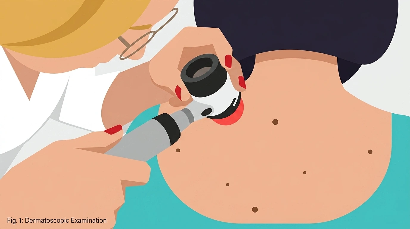
AI tools that read photos of skin lesions and flag possible cancers are already in clinics and phone apps. They were sold as a way to widen access to dermatology, and on lighter skin they often match a specialist. On darker skin they miss things, often enough to change who gets diagnosed in time. The cause is not mysterious, and neither is the fix.
How Big The Gap Is
This isn’t a rounding error. When researchers built the Diverse Dermatology Images dataset to test models across a wide range of skin tones, accuracy dropped by 27 to 36 percent against the models’ usual scores, with the worst results on dark skin and uncommon conditions.
The numbers for individual tools are starker:
- Stanford’s DeepDerm caught malignancies at a sensitivity of 0.69 on lighter skin but only 0.23 on darker skin, close to a three-fold gap in how often it spotted real cancer.
- One melanoma-detection analysis saw sensitivity fall from 67 percent to 11 percent when the same model was pointed at darker skin.
Sensitivity is the number that matters most, because it measures how often the tool catches actual disease. A drop that size means a tool that reliably flags a cancer on pale skin can wave the same cancer through on dark skin.
Why It Fails: The Training Data Is Mostly Light Skin
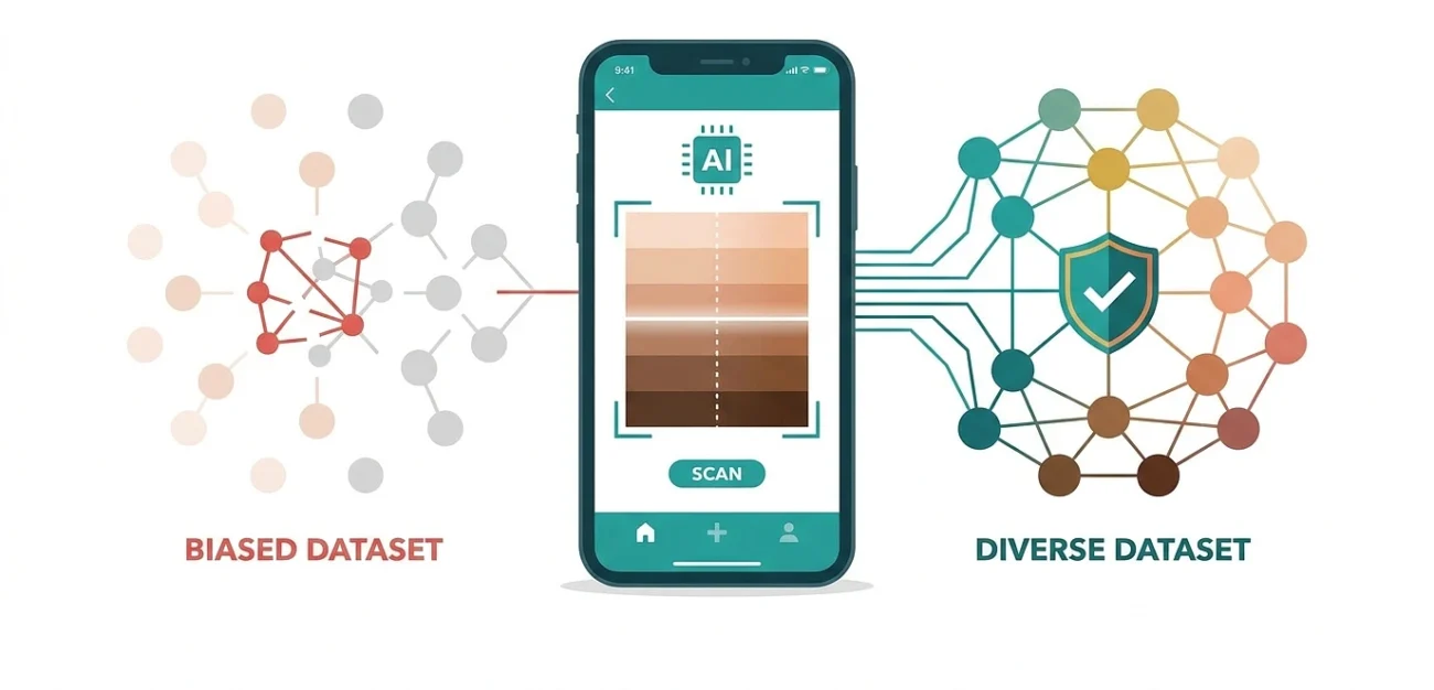
AI learns from the images it’s shown, and those images skew heavily toward one group. The International Skin Imaging Collaboration archive, one of the most widely used training sets in the field, is over 70 percent light-skin images and under 8 percent dark-skin images.
A model trained on that mix gets very good at the patterns it sees thousands of times and stays weak at the ones it barely sees. Nobody designed it to fail darker patients. It never got enough examples to learn them, and the result is a tool that performs like its data, not like the population it’s meant to serve.
Why It Fails: The Diseases Don’t Look The Same On Darker Skin
The data problem is made worse by biology. Many conditions present differently depending on skin tone, so a model that learned the light-skin version of a disease is looking for the wrong signs.
Redness is the clearest example. Inflammation that shows as red on light skin often appears purple or gray on darker skin, so a tool trained to look for red can miss it. Melanoma, eczema, and psoriasis all shift in appearance with pigmentation. This is also why the problem runs deeper than software: the dermatologists who label the training images score worse on dark skin too, so some of the bias is baked in before a model ever trains.
What It Costs Patients
People with darker skin already tend to receive a melanoma diagnosis at a later stage, when it’s harder to treat, and a tool that misses early lesions pushes that the wrong way. With around 3 billion people worldwide lacking reliable access to dermatological care, AI triage is one of the few realistic ways to reach them, so a tool that works on only some skin tones doesn’t close the access gap. It redraws it.
The Data That Fixes It
The fix is known, and it works.
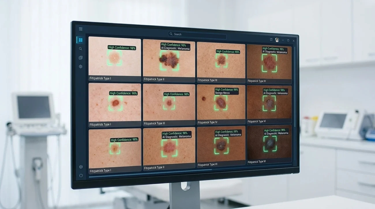
When researchers fine-tuned dermatology models on the diverse DDI images, the performance gap between light and dark skin closed. The retrained models didn’t just improve, they outperformed dermatologists at identifying malignancy on dark-skin images. The lever is the training data, not some unreachable breakthrough.
Two approaches are doing the work:
- Diverse, confirmed datasets. Collecting and pathologically verifying images across the full range of skin tones gives models the examples they were missing. This is the slow, reliable route.
- Synthetic and augmented images. When real dark-skin images are scarce, techniques that realistically adjust skin tone in existing images can expand coverage and lift accuracy on Fitzpatrick IV to VI, without changing the lesion itself.
Neither is exotic. Both come down to showing the model the patients it will see in clinic.
It’s Not Only Dark Versus Light
One nuance keeps getting lost. The fairness conversation tends to split skin into light and dark, but the middle gets forgotten. Fitzpatrick III and IV tones, common across East Asian, Hispanic, and Mediterranean populations, are neither the focus of dark-skin fairness work nor well represented in light-skin datasets. A dataset that corrects only the extremes still leaves a large share of patients underserved.
The Fitzpatrick scale itself is a blunt tool, built to describe how skin reacts to sun rather than to capture the full range of human pigmentation. Better recognition means better data across the whole spectrum, not a single correction at one end.