
Blog
Why Red Bumps Look Different on Darker Skin, and Why AI Keeps Getting It Wrong

Abstract
Walk into most medical training and you learn that inflamed skin is red. A rash is red, an infection is red, an irritated bump is red, and that redness, called erythema, is one of the first things a clinician is taught to look for. The problem is that this lesson was built almost entirely on light skin, and on darker skin inflammation frequently isn’t red at all. It’s purple, or grey, or brown, or barely visible. So the single sign everyone was trained to hunt for is the one sign that often goes missing on the patients who need it seen. And now we’ve handed that same blind spot to machines, which learned from the same lopsided pictures and are repeating the same mistake at scale.
Inflammation On Darker Skin Usually Isn’t Red, It’s Violet Or Grey Or Brown
Start with why the redness disappears, because it’s not that the inflammation is milder, it’s that the color you’re looking for gets masked.
There’s a concept from color theory called simultaneous contrast, and Dr. Jenna Lester, a dermatologist who built one of the country’s few skin-of-color clinics, uses it to explain what’s happening. Color is dynamic, and whatever sits around a color changes how your eye reads it. So when you get erythema, the increased blood flow of inflammation, against a background of melanin-rich skin, it doesn’t hit the eye as red. It reads as violaceous, which is the dermatology word for purplish, or as grey, or dark brown, or sometimes just as an area that’s slightly darker than the skin around it. The inflammation is every bit as real and often just as severe. It simply isn’t wearing the color the textbook promised.
And if your eye hasn’t been trained to see that, you miss it. That’s not a small consequence. When clinicians rely on redness that isn’t there, they underestimate how bad a condition actually is, and the standard severity scoring tools, the ones built around visible erythema, systematically under-rate disease in darker skin. Eczema, psoriasis, rosacea, all of them can look deceptively mild on skin where the redness is masked, right up until the point the disease is advanced. One consequence shows up starkly in the data: eczema hospital admissions have been shown to run up to six times higher in patients of colour, which is what late diagnosis looks like when you count it.
The Bumps Themselves Can Take A Different Shape, Not Just A Different Color
It isn’t only the color that shifts, and this is the part even some clinicians don’t expect, because the actual morphology, the shape and pattern of the bumps, can differ too.
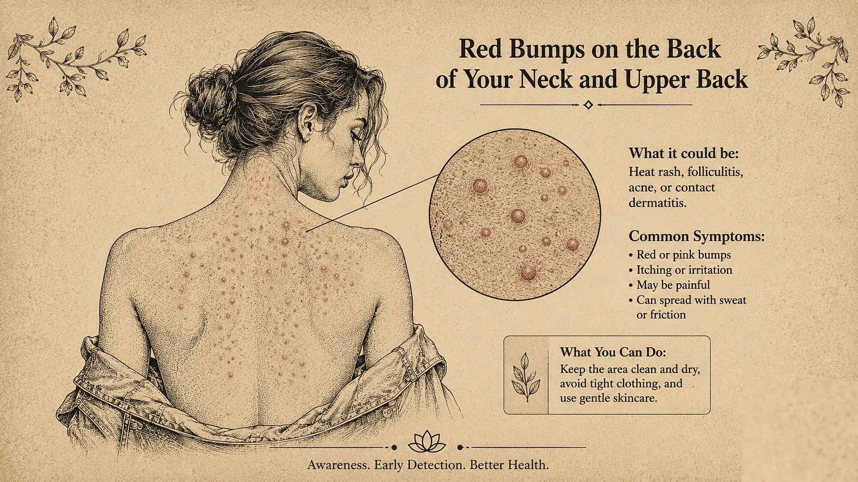
In eczema, for instance, patients of African descent more often show what’s called a follicular or papular pattern, small one-to-two millimetre bumps clustered around the hair follicles on the trunk, the chest, the back, rather than the broad confluent patches in the flexures that the textbooks describe as classic. A clinician scanning for the classic presentation, big red scaly patches behind the knees, can look right at a follicular eczema and not register it, because it doesn’t match the template they were handed. Then there’s the aftermath, because inflammation on darker skin tends to leave post-inflammatory hyperpigmentation, dark marks that linger long after the flare settles, and those marks are their own source of distress and are sometimes the thing that finally brings a patient in, long after the underlying condition should have been caught.
The Reason Both Problems Exist Is The Same Pile Of Pictures
Here's where the two halves of this connect, because the human miss and the machine miss come from one shared root, and it's almost mundane once you see it. It's the images.
Medical students and residents learn to recognize skin disease by looking at photographs, thousands of them, over and over. But audits of dermatology textbooks repeatedly find that only somewhere between four and eighteen percent of those clinical photos show darker skin. So doctors are trained on a nearly monochrome library, they build their pattern-recognition on light skin, and then they carry that pattern to every patient who walks in. Nearly half of new dermatologists have admitted feeling uncomfortable identifying conditions on darker skin tones, which is not a confession of prejudice, it’s a confession about the training set. You get good at recognizing what you’ve seen ten thousand times, and bad at recognizing what you’ve barely seen.
AI Learned Dermatology From The Same Lopsided Library, So It Inherited The Same Blind Spot
Now put a machine through that exact process, because that’s what happened, and the result is depressingly predictable.
AI diagnostic models learn from image datasets, and the big public ones are heavily skewed toward light skin. The widely used ISIC repository runs over seventy percent fair skin, and popular databases were drawn largely from European and Australian populations. Train a model on that, and it learns the features of light skin well and the features of dark skin poorly, and the numbers on what follows are stark. When researchers tested Stanford’s DeepDerm, a celebrated model, on a diverse image set, its sensitivity was around 0.69 on lighter skin and collapsed to 0.23 on darker skin, a nearly threefold gap. Another model, ModelDerm, dropped from 0.41 to 0.12. These are tools that looked highly accurate right up until someone checked whether the accuracy held across skin tones, and it didn’t.
Generative AI has taken the same flaw and started manufacturing it. A 2025 study generated four thousand dermatology images across the major AI models and found that 89.8 percent depicted light skin and only 10.2 percent dark, and separately, only about fifteen percent of the images accurately showed the condition they were supposed to. So the machines aren’t just under-diagnosing darker skin, some of them are now producing a fresh flood of light-skin-dominated teaching images that could train the next generation of doctors and models on the very same imbalance. The blind spot isn’t just being inherited. It’s being reproduced.
What Actually Fixes It Is Boringly Concrete
The encouraging part is that none of this is mysterious or unsolvable, because when researchers have gone looking for the fix, it keeps turning out to be the obvious thing. Show the eye, human or machine, more of what it’s been missing.
When AI datasets were deliberately enriched with diverse skin images, the accuracy gap between light and dark skin narrowed significantly, which tells you the problem was the data all along, not something intractable about darker skin being harder to read. The same logic works on people dermatologists trained deliberately on the full range of presentations get better at spotting the violet, the grey, the follicular bumps, the masked severity. Some practical clinician habits help too, like gently pressing an area to blanch it and reveal subtle underlying inflammation, or simply asking the patient whether their skin has changed color rather than trusting your own read of it. None of that is exotic. It’s just the correction to a training gap that was allowed to run for a very long time, in human eyes first and now in silicon ones, and the fix in both cases is the same. Put the missing pictures back in.